
• Case Provider: Dr. Recep Uzgur
• Solution Used: Eighteeth Helios 500 Intraoral scanner and EXOCAD
• The workflow initiated with scanning using the Helios 500 and file importation into Exocad for further design.
• About the Dentist
Dr. Recep Uzgur was born in Turkey in 1984. After graduating, he completed his PhD in Prosthodontics at Ankara University in 2014. Dr. Recep Uzgur is a key opinion leader for many companies such as Eighteeth, ltero, Dentsply Sirona, Asiga, Osstem Implant, and Bredent lmplant. He is the fellow of the European Prosthodontic Association. He is also a member of various associations, including the Turkish Dental Association, the Turkish Prosthodontics and implantology Association, and the European Prosthodontic Association. Dr, Recep Uzgur's special interests are in esthetic dentistry, digital dentistry, and implantology.
• Case
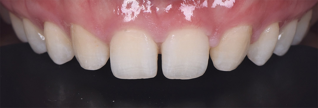
A 33-year-old female patient presented to our clinic with aesthetic concerns, She shared her history of congenital absence of lateral incisors and misalignment for which she underwent orthodontic treatment at another practice, a process she has completed.
The objective of her orthodontic treatment involved moving the canine teeth closer to the central incisors and planning for the spaces between the front four teeth to be closed restoratively post-orthodontics to achieve aesthetic harmony.
• Treatment Planning
During the clinical examination, it was observed that there were spaces between the front four teeth, and considering the canine teeth were sufficiently small, they were considered as potential replacements for the lateral incisors.
The patient expressed a desire for minimal or, if possible, no tooth preparation, so direct composite veneers were initially suggested. However, the patient was interested in ceramic veneers due to their longevity and durability and inauired if the treatment could be done with minimal preparation. Thus, prepless veneers were proposed.
• Treatment Process
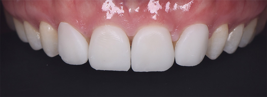
Given that prepless veneers are a challenging treatment and proper indication is critical, and since the primary indications are teeth that require volume on the buccal surface and those with a thick biotype gingiva, it was decided to first create a design and produce temporary veneers from a 3D printer to achieve a real mock-up. This would allow for aesthetic and occlusal checks before obtaining patient approval.


For this, a digital impression was taken using the Helios 500 scanner, and four prepless veneer designs were created in Exocad, followed by the printing of temporary veneers. Temporary veneers were printed by Asiga 3D printer in 45 minutes, These were temporarily bonded to the patient's teeth for aesthetic and occlusal evaluation, giving the patient a preview of the final result before approving the treatment.

• Design and Milling
Folowing this, production ofthe finalveneers commenced using GC LiSi blocks a reinforced ceramic with lithium disilicate that doesn't require crystallization and can be finalized with mechanical polishing, allowing for adhesive cementation without heat treatment when color match is achieved.
Since patient approval was taken, same digital design was used for final veneers. No crystallization was required, and just mechanical polishing were applied to veneers, For milling Redon GTR unit was emploved.

• Adhesive Cementation
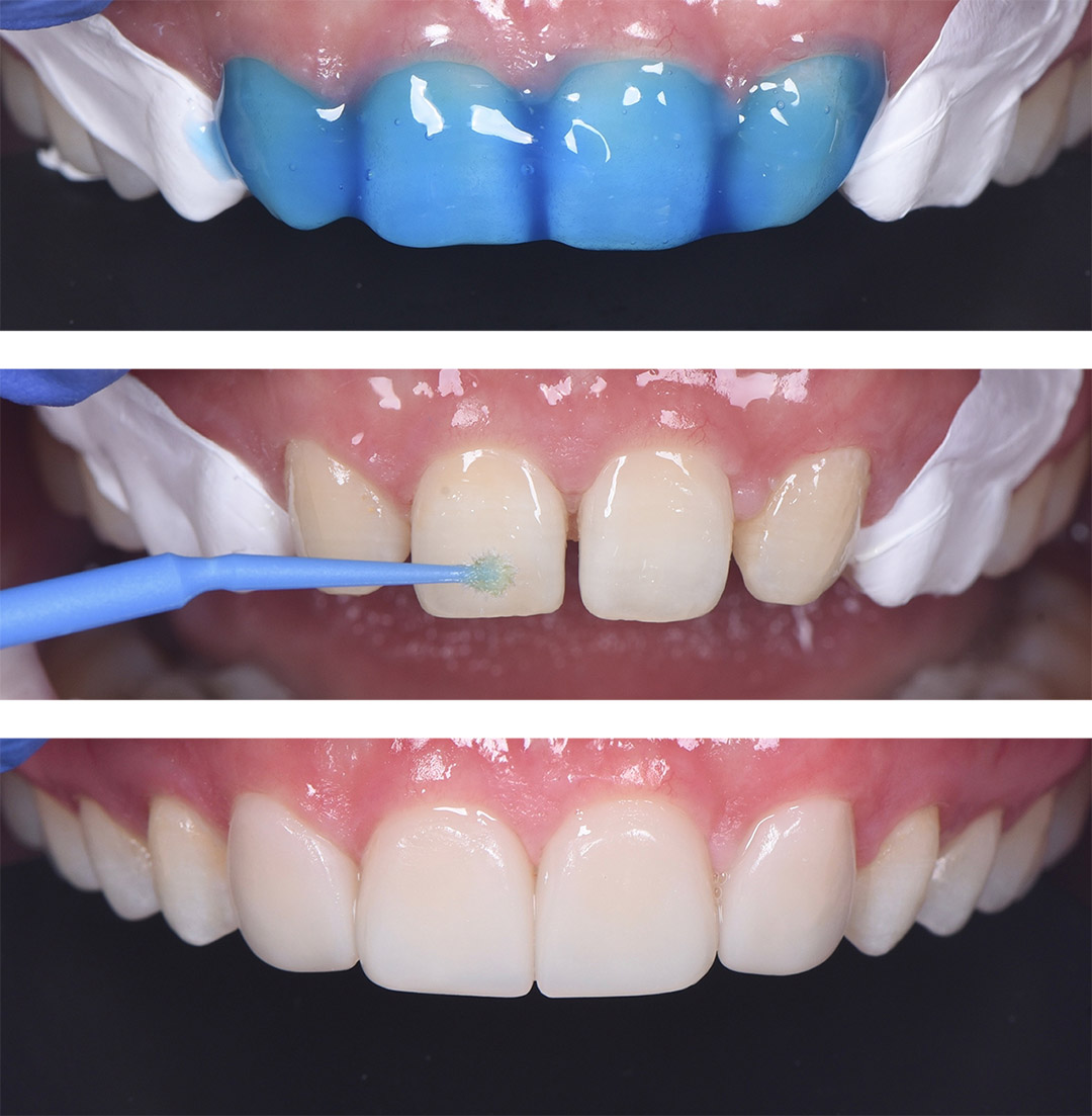
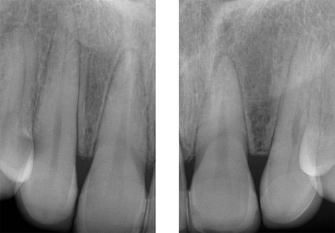
After the production and polishing phase, the final prepless veneers were tested in th " atient's mouth with trial paste and shown to the patient. The final phase involved cementing the veneers in place with Variolink LC adhesive cement using a total etch bonding technigue. Remarkably, this entire procedure was completed without touching the anterior four teeth and without the need for local anesthesia.

• Intraoral Scanning Protocol
A standard scanning protocol was meticulously followed. First upper arch digital scan then lower arch scan were completed. Buccal scans were conducted on both sides to enhance the accuracy of the impression.
The advanced artificial intelligence of the Helios 500 scanner operated in the background, optimizing buccal scanning stage by correcting any potential errors that could arise from operator or patient movements.

• Verification with Periapical X-Ray


• Comment of Dentist
For this particular treatment, the Helios 500 intraoral scanner was employed, ensuring a streamlined and entirely digital workflow.
The scanner efficiently captured detailed images of each jaw in under a minute for both sides.
In cases involving prepless veneers, the accuracy of scanning and miling is critical, which is why the Helios 500 intraoral scanner plaved a significant role. Despite the challenges, the Helios 500 completed this case flawlessly.
Thanks to the Helios 500's capabilities in managing such a treatment, the case was brought to a successful conclusionwithin 6 hours.